Machine Learning Research
Machine Learning
Registration
The analysis of multimodal data at multiple time points is key in order to generate disease and treatment prediction models. Thus we must align patient SD-OCT scans, as well as extracted disease features, into a common reference frame. In order to perform this registration, certain landmark features are required. These landmarks, obtained from both SD-OCT and color fundus (CF) images, include the retinal vasculature, the fovea position as the functional center of vision, and the optic nerve head connecting the eye to the brain.
Fovea detection
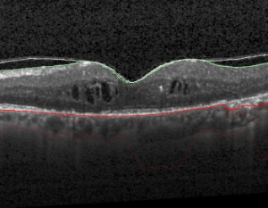
The retinal fovea is the location of the highest visual acuity and is present in all patients, thus it is critical to vision and highly suitable for use as a primary landmark for cross-vendor/cross-patient registration for precise comparison of disease states. However, the location of the fovea in diseased eyes is extremely challenging to locate due to varying appearance and the presence of retinal layer destroying pathology. Thus categorizing and detecting the fovea type is an important prior stage to automatically computing the fovea position. We developed an automated cross-vendor method for fovea distinction in 3D SD-OCT scans of patients suffering from RVO, categorizing scans into three distinct types (see figure below).
“Automated retinal fovea type distinction in spectral-domain optical coherence tomography of retinal vein occlusion.” SPIE Medical Imaging.
Wu, Jing, et al.
International Society for Optics and Photonics, 2015.
OCT Vessel Segmentation
We developed a fully automated cross-vendor method to acquire retina vessel locations for OCT registration from fovea centered 3D SD-OCT scans based on vessel shadows. Extracted RPE layers are combined to generate a projection image featuring all candidate vessel shadows, as illustrated in the figure above. Image processing methods are then applied to remove false positive shadow regions such as those caused by exudates and cysts. Validation of segmented vessel shadows uses ground truth vessel shadow regions identified by expert graders at the Vienna Reading Center (VRC).
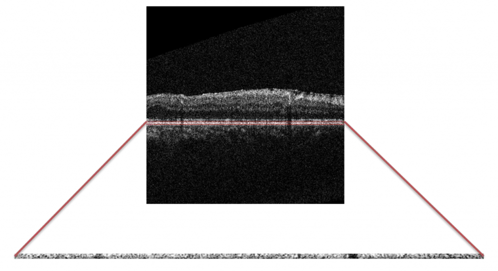
“Automated vessel shadow segmentation of fovea-centered spectral-domain images from multiple OCT devices.” SPIE Medical Imaging.
Wu, Jing, et al.
International Society for Optics and Photonics, 2014.
CF Vessel Segmentation

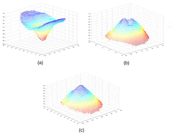
We also developed an algorithm to segment retinal vessels in color fundus images. In the figure above, (a) denotes the original image, (b) the ground truth segmentation, and (c) the vessel segmentation of our algorithm.
“Rotation invariant eigenvessels and auto-context for retinal vessel detection.” SPIE Medical Imaging.
Montuoro, Alessio, et al.
International Society for Optics and Photonics, 2015.
Registration using Vessels
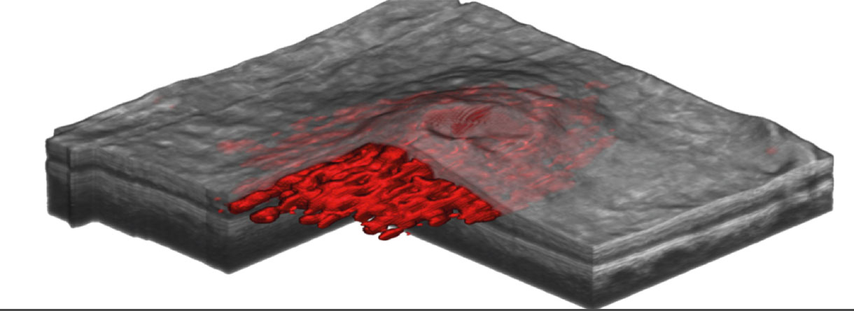
We developed a multiple scanner vendor registration method for pathological retinal 3D spectral domain optical coherence tomography volumes based on our automated vessel shadow segmentation. The figure above illustrates the segmented vessels in red between the green ILM and blue RPE surface. Myronenko’s Coherent Point Drift is applied to the segmented retinal vessel point sets in order to generate the registration parameters required.
“Stable registration of pathological 3D-oct scans using retinal vessels.”
Wu, Jing, et al.
Ophthalmic Medical Image Analysis First International Workshop, OMIA 2014.