Machine Learning Research
Machine Learning
Segmentation
In order to optimize the treatment of patients suffering from various eye diseases, the interpretation of SD-OCT scans is important. Therefore it is necessary to know which image structures in the scans are relevant in terms of the disease and disease progression and to be able to locate and extract these pathologies automatically and reproducibly in a wide range of scan qualities, as manual annotation is a time-consuming and complex procedure. For example, these automatic segmentations can then be used for prediction of visual acuity or optimal time span between treatments.
Layer Segmentation
Obviously, the retinal layers are relevant image structures for the purpose of interpreting OCT scans. Our collaboration partners from Iowa developed the Iowa Reference Algorithm, a graph-theoretic segmentation method for the simultaneous segmentation of multiple 3-D surfaces, where segmentation results for the retinal layers are visualized in the figure on the right.
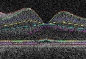
“Automated 3-D Intraretinal Layer Segmentation of Macular Spectral-Domain Optical Coherence Tomography Images.”
Garvin MK, Abramoff MD, Wu X, Burns TK, Russell SR, Sonka M.
IEEE Trans Med. Imaging, 28, 9, 1436-47, 2009.
Vitreous Segmentation
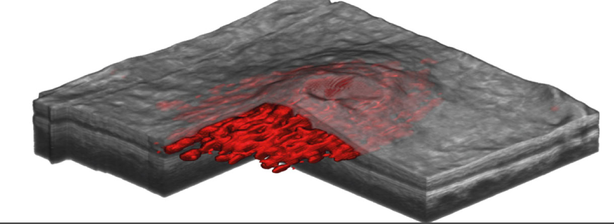
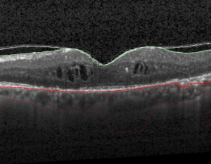
Precise and reproducible detection, quantification and classification of the posterior vitreous boundary and its adhesions at the macula is of major importance. For instance, efficacy of pharmacologic treatment of vitreomacular interface disorders depends on strictly defined characteristics of the vitreous adhesion. We developed a method to fully automatically segment the vitreous boundary in SD-OCT scans, where a segmentation result is illustrated in the figure on the right.
“Automatic segmentation of the posterior vitreous boundary in retinal optical coherence tomography.”
Montuoro, Alessio, et al.
Investigative Ophthalmology & Visual Science 56.7 (2015): 5275-5275.
IRC and SRF Segmentation
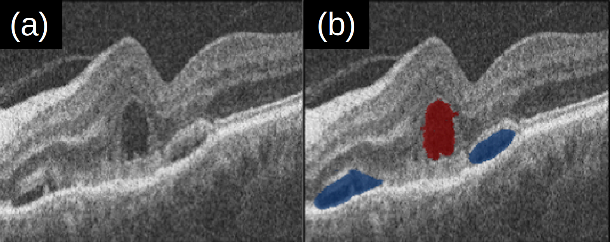
As intraretinal cystoid fluid (IRC) and subretinal fluid (SRF) are well-known characteristics of retinal vascular disease, we developed an automatic segmentation algorithm for IRC and SRF, based on deep learning. Results are visualized in the figure on the right, where (a) shows the original, and (b) the segmented scan (red=IRC, blue=SRF).
„Predicting Semantic Descriptions from Medical Images with Convolutional Neural Networks.”
T. Schlegl, S. M. Waldstein, W.-D. Vogl, U. Schmidt-Erfurth, G. Langs (2015).
Information Processing in Medical Imaging, IPMI 2015.
Unsupervised OCT Segmentation
Currently, not all structures are enlightened and interpretable. One objective of this project is to find an appropriate categorization of image structures and to correlate these found structures with the disease. This is a difficult task, because medical images show high variability in appearance patterns, which can be distinctive of different pathologies, as well as being heavily affected by noise and acquisition artefacts. We plan to use deep learning to detect image biomarkers automatically, in an unsupervised fashion.