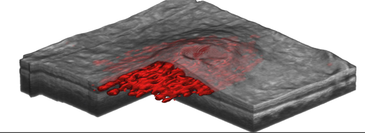
Machine Learning Research
Machine Learning
Preprocessing
While OCT Scans offer a variety of advantages (among others non-invasive scanning, µm resolution and fast acquisition times) they also present some challenges for automated processing.
Motion Correction
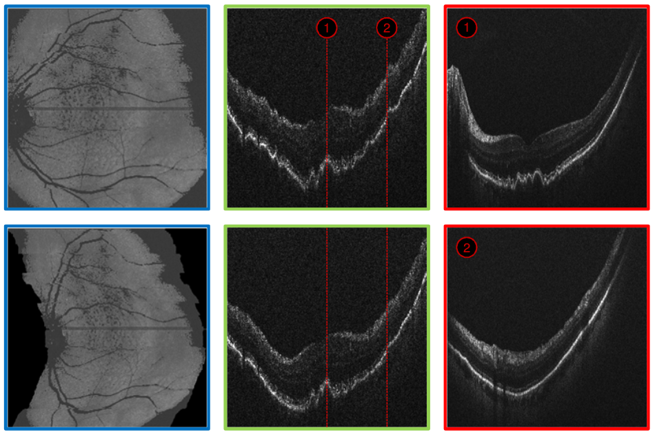
Retinal OCT scans are generated by scanning through the retina generating one column at a time. These scanned columns are then assembled first to 2D slices and then to 3D volumes. Even though this process is rather fast (in the range of 5 seconds) due to the high resolution (typically around 5µm per voxel in axial scanning direction) even tiny involuntary eye movements are visible as scanning artefacts. These artefacts are visible as discontinuities in the vascular structures and ripples in the retinal layers (see first row of image below).
We developed a method for correcting these artefacts while preserving the shape of potential pathologies (see second row of image below).
“Motion artefact correction in retinal optical coherence tomography using local symmetry.”
Montuoro, Alessio, et al.
Medical Image Computing and Computer-Assisted Intervention–MICCAI 2014. Springer International Publishing, 2014. 130-137.
Denoising
Another challenge present in OCT scans is the rather low signal to noise ratio. This can be addressed during the acquisition by scanning the same spatial location multiple times and averaging the result (called oversampling). However this requires special eye tracking hardware and increases the overall scanning time. Another approach is to remove the noise after the acquisition using software algorithms. Such algorithms have to be designed and validated very carefully in order to only remove the noise from the scan without introducing any new artefacts (such as blurring tissue boundaries).
We developed a method based geodesic denoising and validated it on real pathological OCT scans and synthetically generated noise-free OCT volumes. This Video shows an OCT scan before (left) and after (right) noise suppression.
“Geodesic denoising for optical coherence tomography images.” SPIE Medical Imaging.
Varnousfaderani, Ehsan Shahrian, et al.
International Society for Optics and Photonics, 2016.
“Improve synthetic retinal OCT images with present of pathologies and textural information.” SPIE Medical Imaging.
Varnousfaderani, Ehsan Shahrian, et al.
International Society for Optics and Photonics, 2016.